Surgical repair of symblepharon — conjunctival adhesions from chemical burns, Stevens-Johnson syndrome, and cicatrizing conjunctivitis — with mucous membrane and amniotic membrane grafting.
Medically reviewed by EyePlastics Medical Editorial BoardASOPRS oculoplastic surgeonsLast updated June 2026
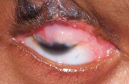
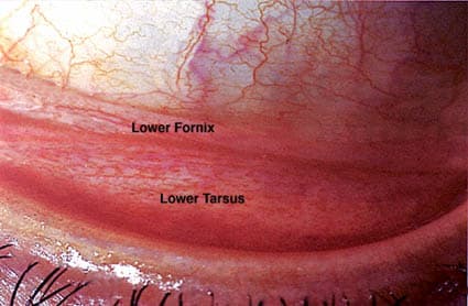
Symblepharon is an adhesion between the palpebral conjunctiva (lining the inner eyelid) and the bulbar conjunctiva (covering the eye surface). These bands of scar tissue tether the eyelid to the eyeball, restricting eyelid movement, limiting ocular motility, and distorting the conjunctival fornix — the recessed pocket where the eyelid lining meets the eye. In severe cases the fornix may be obliterated entirely and the eyelid can fuse to the globe (ankyloblepharon); accompanying corneal surface failure (keratinization, limbal stem cell deficiency, opacification) can lead to severe vision loss.
Symblepharon is most commonly caused by severe chemical injury to the eye. Related conditions include Eyelid Laxity and Lagophthalmos, which may coexist when eyelid scarring accompanies symblepharon.
Symblepharon results from any condition that damages both the palpebral and bulbar conjunctival surfaces simultaneously, allowing them to fuse during healing:
Symblepharon severity is graded by the degree of fornix obliteration:
Associated findings include keratinization of the conjunctiva, limbal stem cell deficiency (LSCD) causing corneal vascularization and opacification, and trichiasis from distorted lid margin.
The goal in the acute phase of chemical injury or SJS is to prevent symblepharon formation:
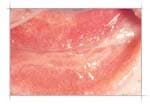
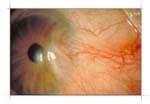
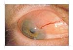
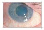
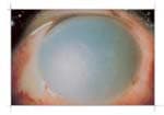
Once symblepharon has matured, surgical lysis and fornix reconstruction are required to restore motility and eyelid function. Surgery is indicated for:
After surgical lysis of the symblepharon bands, the raw surfaces must be covered with a graft to prevent re-adhesion. Autologous mucous membrane (from buccal mucosa, nasal septum, or hard palate) is harvested and sutured to line the reconstructed fornix. MMG provides a durable, non-keratinizing surface.
Amniotic membrane can be used as an adjunct or alternative to autologous mucous membrane for fornix reconstruction. It provides anti-inflammatory, anti-fibrotic, and anti-angiogenic properties, promoting re-epithelialization. Often combined with a conformer to maintain fornix depth post-operatively.
When symblepharon is accompanied by limbal stem cell deficiency — evidenced by corneal vascularization, conjunctivalization of the cornea, and recurrent epithelial breakdown — limbal stem cell transplantation (LSCT) is necessary before or concurrent with symblepharon repair to restore a stable corneal surface. Sources include:
A conformer or custom ocular prosthesis is placed immediately after fornix reconstruction and maintained to prevent re-adhesion during healing. In progressive cicatrizing conditions (OCP), the conformer may need to be worn indefinitely.
In autoimmune cicatrizing conjunctivitis (OCP, SJS), treatment of the underlying systemic disease is essential to halt progression:
Oculoplastic management of symblepharon is most effective when performed in collaboration with rheumatology, dermatology, or immunology for systemic disease control.
Connect with a board-certified oculoplastic surgeon who specializes in symblepharon.
Search the Directory →Oculoplastic Surgery — Patient Education
Lagophthalmos